Welcome to
Together is a new resource for anyone affected by pediatric cancer - patients and their parents, family members, and friends.
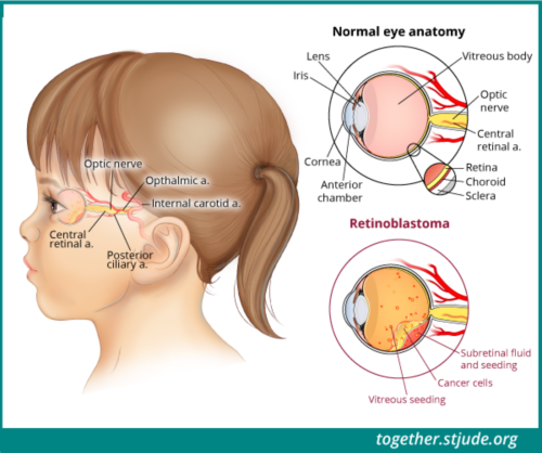
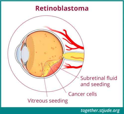
Learn MoreRetinoblastoma is a cancer of the eye. It most often occurs in young children, usually before 3 years of age. This cancer rarely develops in children older than age 5. Retinoblastoma forms in the retina of the eye. The retina is a thin layer of nerve tissue in the back of the eye. The cells of the retina detect light and color. Retinoblastoma may affect one eye (unilateral, most common) or both eyes (bilateral).
Retinoblastoma is a cancer that forms in the retina of the eye. The retina is a thin layer of nerve tissue in the back of the eye.
Retinoblastoma is rare. About 250-300 children are diagnosed per year in the United States. Retinoblastoma can be hereditary (able to pass down in families) or non-hereditary.
In most patients, retinoblastoma is confined to the eye, and the cancer is very curable. The cure rate for this cancer is above 95% because of early detection and available treatments. If the disease is not treated, it can spread to other parts of the body, where it becomes much harder to treat. Patients with hereditary retinoblastoma are also at higher risk for other cancers later in life.
Treatments for retinoblastoma include chemotherapy to shrink the tumor, locally delivered therapy (anti-cancer agents given directly to the eye), focal therapy (laser or freezing therapy delivered directly to the eye), surgery to remove the eye, and radiation therapy.
In most cases, retinoblastoma results from a change or mutation in a gene known as the retinoblastoma (RB1) gene. When there is a mutation of the RB1 gene, cells grow and divide abnormally.
There are two types of retinoblastoma: non-hereditary (sporadic) and hereditary.
The most common form of retinoblastoma is sporadic, or non-hereditary. Sporadic retinoblastoma is not passed down in families. There is a change in the RB1 gene in a single cell in the retina of one eye. These cells divide and form a tumor. Most patients with only one eye affected (unilateral) have sporadic retinoblastoma.
About 25-30% of children with retinoblastoma have the hereditary form of the condition. Hereditary retinoblastoma is the result of a change (mutation) in the RB1 gene in every cell in the body. Most patients with hereditary retinoblastoma have both eyes affected (bilateral). Approximately 10-15% of patients with unilateral retinoblastoma and all patients with bilateral retinoblastoma have the hereditary form of the disease. Children with hereditary retinoblastoma are at higher risk for developing other cancers later in life. Children with hereditary retinoblastoma may be the first in their families with this gene mutation. They then have a 50% chance of passing the gene on to their children.
Genetic testing is important in retinoblastoma. The type of cancer — sporadic or hereditary — can affect risk of future cancers and may influence future life decisions.
A doctor may find retinoblastoma during a routine eye exam. However, many times a parent or relative first notices that a child’s eye looks different from normal. Some of the symptoms of retinoblastoma are common to other eye problems, so it is important to see a pediatric eye doctor for evaluation.
Signs and symptoms of retinoblastoma include:
If a child shows signs of retinoblastoma, it is important to see a pediatric ophthalmologist (eye doctor for children). An eye doctor may do an office screening exam to see if further testing is needed.
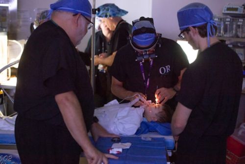
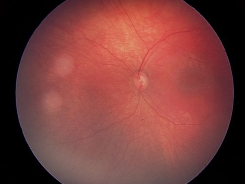
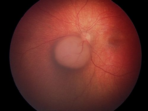
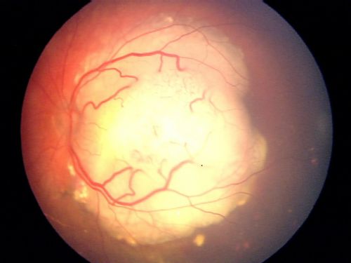
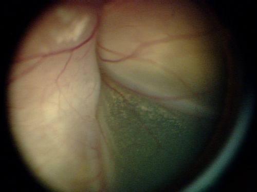
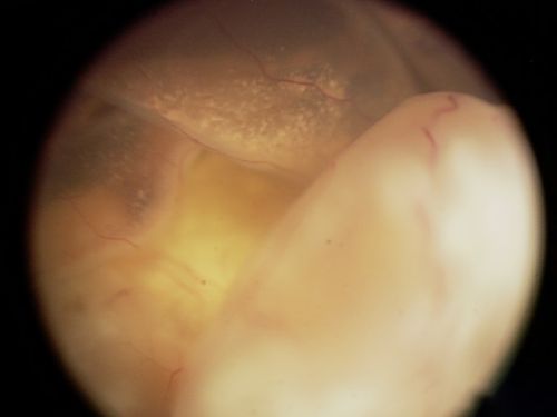
Eye Exam Under Anesthesia - A diagnosis of retinoblastoma is made using an eye exam under anesthesia (EUA). In this test, a doctor examines the eye closely using special lights and a magnifying lens after the eyes are dilated. This procedure is done while the child is asleep so that the doctor can view the entire retina. A special camera takes digital pictures of the eye and tumor.
A biopsy is not used to diagnosis retinoblastoma because this might cause the cancer cells to spread.
Other types of tests may be used to help diagnose and determine the extent of retinoblastoma. These tests include:
If there are concerns that the disease has spread outside the eye, doctors will take samples of the bone marrow and cerebrospinal fluid. Additional imaging tests may be performed to look at the brain and spinal cord.
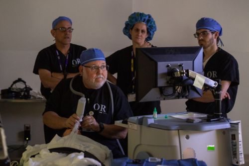
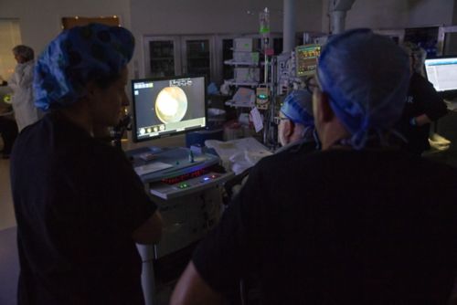
An exam under anesthesia (EUA) allows the doctor to examine the eye closely using special lights and a magnifying lens.
If retinoblastoma is not treated, it can spread:
Large tumors may break into smaller tumors that float within the eye fluid. These are called vitreous seeds.
Tumors of the eye are “grouped” or classified from Group A to Group E. In the past, retinoblastoma tumors were classified using a numbered grouping system (Group I to Group V), called the Reese-Ellsworth classification.
Grouping describes the likelihood of saving the eye. The group is based on tumor size, location, and extent of spread inside the eye. If both eyes are affected, each eye is graded separately.
Group A tumors are small, and they are located away from the center of vision. Patients with Group A tumors have a good chance of maintaining normal eye function and vision during and after treatment of the tumor.
Group E tumors are large and have broken apart inside the eye (vitreous seeding). These tumors are more likely to spread outside the eye. Children with Group E tumors are expected to have reduced or complete loss of vision, and there is a lower chance to be able to keep the eye, even with treatment.
Tumor Group | Risk of Losing the Eye | Clinical Features |
---|---|---|
A | Very low risk | Small tumor located only in the retina; not near important structures |
B | Low risk | Larger tumor and/or located near important structures |
C | Moderate risk | Tumor is mostly well-defined with small amounts of spread or seeding |
D | High risk | Tumor is large or poorly defined with high amount of seeding |
E | Very high risk | Very large tumor that is affecting eye structure and function; higher chance of spread |
The stage of cancer refers to whether it has spread outside the eye. Stage I and Stage II mean the tumor is located only within the eye. Stages III-IV indicate the tumor has spread to local tissues surrounding the eye or to distant sites (metastatic disease).
The tumor is small and located only in the retina. It is not near important eye structures.
The tumor is larger, or it may be located near important eye structures.
The tumor is mostly well-defined but has small amounts of spread or seeding.
The tumor is large or poorly defined and has a high amount of seeding.
The tumor is very large and affects eye structure and function. There is a higher chance of spread.
Most children with retinoblastoma can be cured. The main factor that influences a child’s survival is whether the tumor has spread outside of the eye. If it is localized to the eye, the prognosis for patient survival is excellent. If the cancer has spread to other parts of the body, the illness becomes harder to treat.
Several factors influence the outlook for a patient’s treatment and recovery:
Patients with cancer confined to the eye have survival rates >95% with treatment. If the cancer has spread to the eye socket (orbit), lymph nodes, bone marrow, bones, or liver, patients have survival rates of about 80% with intensive chemotherapy, autologous stem cell rescue, and radiation therapy. When cancer has spread to the brain or cerebrospinal fluid at the time of diagnosis, the prognosis is usually poor (<10% survival).
In the treatment of retinoblastoma, doctors and families must consider how the disease is affecting the child. The focus is first on cure and then on preserving the eye with as much vision as possible. The most important factor for saving the eye is the extent of disease at diagnosis. If there is concern about spread of the disease or if treatment is unlikely to preserve meaningful vision, doctors may recommend surgical removal of the eye (enucleation).
In treating retinoblastoma, patient survival is not always the same thing as “eye survival.” The group or classification of the tumor affects whether the eye can be saved. When tumors are grouped A – C, the eye and vision are easier to save. Tumors that have broken apart inside the eye with vitreous seeding (Group D or E) are hardest to treat.
When tumors are in both eyes (bilateral retinoblastoma), usually an attempt is made to save both eyes and preserve as much vision as possible. This is successful in about 70-85% of cases.
More advanced disease in unilateral retinoblastoma (group D or E) may require removal of the diseased eye. If the eye is removed, doctors will examine tissue samples under a microscope to make sure that retinoblastoma has not spread outside the eye. Some patients at high risk for spread outside the eye need chemotherapy. Most patients with unilateral retinoblastoma who have surgical removal of the eye will not need any additional treatment. However, careful monitoring of the remaining healthy eye is essential.
The therapies used to treat retinoblastoma depend on whether the tumor affects one eye (unilateral) or both eyes (bilateral), the extent of disease within the eye, and whether the tumor has spread to other places in and around the eye. Doctors will also consider other factors including:
Surgery, focal therapy, chemotherapy and/or radiation therapy are used to treat retinoblastoma. Usually, a combination of treatments is used. Before starting therapy, the treatment plan should be discussed with the patient and family in detail to outline risks and benefits, both short- and long- term.
Very rarely, retinoblastoma spreads to the brain or cerebrospinal fluid. It can also spread to bones, bone marrow, lymph nodes, or liver. In these cases, intensive chemotherapy may be needed, followed by autologous bone marrow transplant. Some patients may also need radiation therapy.
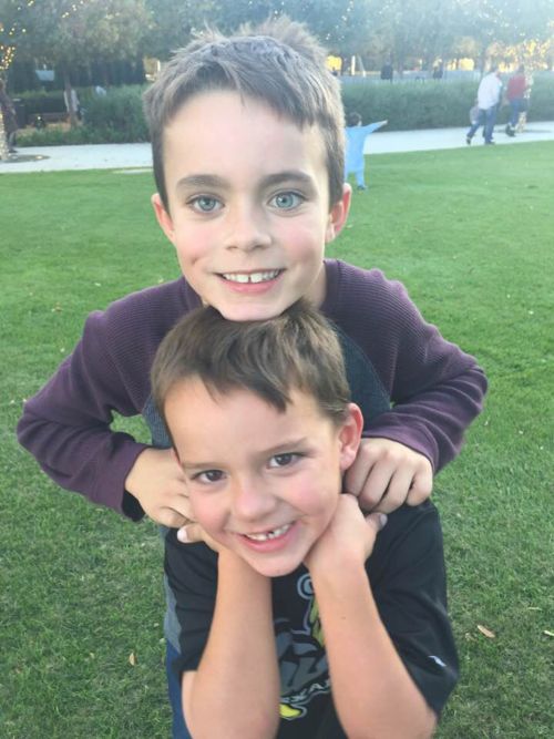
Hunter was diagnosed with retinoblastoma at 11 months of age. He was treated with surgical removal of his eye and has been cancer free since. Hunter enjoys all sports and is a great big brother.
Having only one eye really doesn’t affect what Hunter can do. His biggest physical challenge is making sure he doesn't knock his drink over each day. But, basketball, baseball, golf? It doesn’t really affect him. He might have to make a few adjustments: change his stance, make sure to turn his head. But, everyone has limitations to overcome.
Hunter (top), age 7, with brother Jake
Children will receive regular exams including eye exams under anesthesia (EUA) throughout treatment and for a period of time after treatment. Retinoblastoma recurrences can be managed with focal therapy if caught early. The frequency and duration of follow-up care are determined by the child’s medical team.
Lifelong follow-up for eye health is important for all survivors of retinoblastoma. Eye exams every 6-12 months (depending on age) by an ophthalmologist are recommended as part of routine health care. Protective eyeglasses must also be worn to prevent eye injury.
Patients with eyesight loss may need glasses or other vision assistance. A low vision specialist can help patients who have significant eyesight loss, and early introduction to braille can be helpful.
When an eye is removed surgically (enucleation), the surgeon will place an orbital implant at the time of the initial surgery. After the eye socket has healed, the child can be fitted for a prosthetic or artificial eye. The prosthetic eye is customized for the patient to make the eye look and feel as natural as possible. Most children who need their eye removed have already adapted to changes in vision before surgery. Visual behaviors do not usually change significantly after eye removal. Children with a prosthetic eye can do their normal activities including sports and swimming.
Good care of the prosthetic eye is important. An ocularist is a person who is trained to make and care for ocular prostheses. The prosthetic eye is adjusted as the child grows, usually every 6 months. Regular exams every 6-12 months by an ocularist are recommended for patients with a prosthetic eye. These visits involve cleaning the prosthetic eye, making sure the fit is correct, and addressing any problems.
Eye problems depend on the type of treatment the patient received. Some of the more common eye problems after retinoblastoma include:
Patients treated with external beam radiation therapy to the eye are at risk for certain complications, or late effects of therapy. These may include the following problems:
Survivors of hereditary retinoblastoma have a greater risk of developing other cancers later in life. Young patients are followed with routine brain MRIs until the age of 5 to screen for pineal gland tumors. After age 5, there are no standard recommendations for routine imaging to screen for second cancers. Patients should tell their primary care providers about their medical history and have yearly physician visits (at a minimum) to monitor health.
For general health and disease prevention, all cancer survivors should adopt healthy lifestyle habits. It is important to have regular checkups and screenings by a primary care physician and have a personalized survivorship care plan. Survivors who were treated with systemic chemotherapy or radiation should be monitored for late effects of therapy.
—
Reviewed: September 2019