Skip to Main Content

Welcome to

Together is a new resource for anyone affected by pediatric cancer - patients and their parents, family members, and friends.

Learn More
Blog

Ependymoma

What is ependymoma?

Ependymoma is a tumor of the brain and spinal cord. About 200 children are diagnosed with ependymoma in the United States each year.

Ependymomas are most common in children under age 5. But these tumors may happen at any age. Rarely, they can spread through the cerebrospinal fluid (CSF). This fluid surrounds and protects the brain and spinal cord.

Ependymomas grow from cells that line the fluid-filled ventricles of the brain and central canal of the spinal cord.

Ependymomas grow from cells that line the fluid-filled ventricles of the brain and central canal of the spinal cord.

In children, about 75% of ependymomas are in the posterior fossa region of the brain. This space near the base of the skull contains the cerebellum and brain stem. But ependymomas can also form in other areas of the central nervous system (CNS).

The parts of the CNS that are affected depend on the tumor’s location. These include the:

  • Cerebrum
  • Cerebellum
  • Brainstem
  • Spinal cord

Many ependymomas form in the 4th ventricle in the posterior fossa. These tumors are most likely to affect the brain stem and cerebellum.

Treatment for ependymoma includes:

  • Surgery to remove as much of the tumor as possible
  • Radiation therapy to kill any remaining cancer cells
  • Chemotherapy is sometimes used before a second surgery. Chemotherapy is not typically used if the entire tumor has been removed.

The survival rate for childhood ependymoma is 50–70% (5–7 of 10). Prognosis depends on:

  • The location of the tumor
  • The amount of tumor that can be removed with surgery
  • Molecular features (the underlying biology) of the tumor

Doctors are learning more about the biology and molecular features of ependymoma. These details can help predict how a tumor might respond to treatment. Ependymoma has many molecular subtypes with different clinical features.

Ependymoma can come back after treatment. Children often need long-term care to watch for this.

Symptoms of ependymoma

Signs and symptoms of childhood ependymoma depend on several factors. These include the tumor's size and location and the child’s age and stage of development.

Ependymoma symptoms may include:

  • Headache
  • Nausea and vomiting, often worse in the morning
  • Back or neck pain
  • Vision problems
  • Loss of balance or problems walking
  • Leg weakness
  • Seizures
  • Irritability or confusion
  • Problems urinating (peeing)
  • Change in bowel function
  • Increased head size in infants

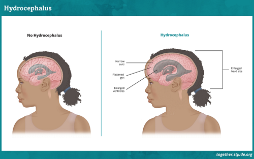
As the tumor grows, it may block the normal flow of CSF. This causes a fluid buildup in the brain known as hydrocephalus. The fluid increases pressure in the brain. Many symptoms of ependymoma are due to hydrocephalus.

Sometimes an ependymoma can block the normal flow of cerebrospinal fluid (CSF). Hydrocephalus occurs when too much CSF builds up in the ventricles.

Sometimes an ependymoma can block the normal flow of cerebrospinal fluid (CSF). Hydrocephalus occurs when too much CSF builds up in the ventricles.

Risk factors for ependymoma

Most ependymomas occur in infants and children under age 5.

Certain changes in genes and chromosomes in the tumor cells are linked to ependymoma. These are not usually passed down from parents. In most cases, it is not known why these genetic changes occur.

Some children may have a higher risk for ependymoma due to neurofibromatosis type 2 (NF2). This rare condition is passed through families.

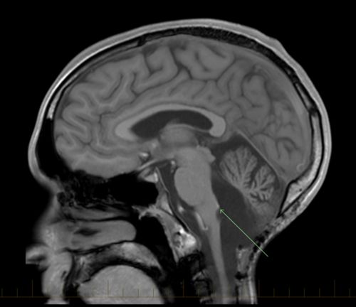
Many ependymomas form in the fourth ventricle in the posterior fossa region. Tumors in this location are most likely to affect the brain stem and cerebellum.

Many ependymomas form in the 4th ventricle in the posterior fossa region. Tumors in this location are most likely to affect the brain stem and cerebellum.

Diagnosis of ependymoma

Doctors use these tests to diagnose ependymoma:

  • Physical exam and medical history
  • Neurological exam to check brain, spinal cord, and nerve function
  • Imaging tests such as a magnetic resonance imaging (MRI) of the brain and spinal cord
  • Lumbar puncture to look for cancer cells in the cerebrospinal fluid (CSF)
  • Biopsy to look for cancer cells in brain tissue

Types of ependymoma

Ependymomas can have different names based on:

  • Their location in the brain or spine
  • The tumor features
  • Genetic changes (mutations) that caused the tumor

Since there are different types of ependymomas, your child's tumor needs molecular testing for an accurate diagnosis.

Common types of ependymoma in children include:

  • Posterior fossa ependymoma
    • Posterior fossa ependymoma group A (PFA)
    • Posterior fossa ependymoma group B (PFB)
    • Posterior fossa ependymoma, not otherwise specified (NOS)
  • Supratentorial ependymoma
    • Supratentorial ependymoma, ZFTA fusion, (formerly called RELA ependymoma)
      • Most common is the ZFTA-RELA fusion, but ZFTA can have different fusion partners, such as MAML or NCOA.
    • Supratentorial ependymoma, YAP1 fusion
    • Supratentorial ependymoma, not otherwise specified (NOS)
  • Spinal ependymoma
  • Myxopapillary ependymoma
  • Spinal ependymoma, MYCN amplified

Molecular testing on your child’s tumor is crucial. This will help the doctor create the best treatment plan for your child.

Treatment of ependymoma

Treatment of ependymoma usually includes surgery followed by radiation therapy. Some patients may also get chemotherapy.

Treatment depends on:

  • The type and location of the tumor
  • Age of the child
  • If the tumor is newly diagnosed or has returned after treatment

Risk-based approaches to treatment classify patients based on tumor features and predicted outcomes. Doctors consider:

  • Whether surgery completely removed the tumor
  • Whether the disease has spread through the body
  • How the cells look under a microscope (histology)
  • Genetic features of the tumor

Drain or shunt

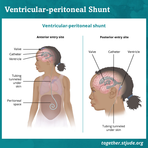
Some children with hydrocephalus due to ependymoma may have external drains or shunts placed in the brain to prevent CSF from building up. 

A shunt is a small tube that drains CSF to prevent a build-up of fluid.

A shunt is a small tube that drains CSF to prevent a build-up of fluid.

Prognosis for ependymoma

The overall 10-year survival rate for pediatric ependymoma is about 50–70% (5–7 of 10) in the United States.

Factors that affect the chance of cure include:

  • Location of the tumor
  • Type and grade of the tumor
  • If the cancer has spread to other parts of the body
  • If surgery can completely remove the tumor
  • Age at diagnosis
  • If the cancer has come back after treatment
  • Molecular features of the tumor

Children with certain types of ependymoma tend to have a better chance for long-term survival. But late recurrences or relapse (beyond 5 years) may occur.

Ependymoma is hard to cure without successful surgery to remove the tumor. If surgery is incomplete or ependymoma recurs, the long-term prognosis is usually poor.

Prognosis for ependymoma depends on many factors. Every case is different. Always talk to your care team about your child’s case.

Support for patients with ependymoma

Palliative care and support services such as rehabilitation, psychology, and social work can help ependymoma patients and families manage symptoms, promote quality of life, and make care decisions. Talk to your care team about what problems to expect and ways to help manage them.

Patients need ongoing follow-up care, lab tests, and routine imaging to watch for disease recurrence and other medical problems. The care team will set a schedule based on the tumor type, response to treatment, and individual patient needs.

General follow-up schedule for ependymoma survivors:

  • Years 0–3: MRI of the brain every 3–4 months
  • Years 3–5: MRI of the brain every 6 months
  • After 5 years: MRI of the brain every 12 months for 2–5 more years

Survivors are at risk for problems such as:

These problems may be caused by the tumor or they may be long-term or late effects related to treatment. Regular checkups and screenings by a primary care physician are important to watch for health problems that can develop years after treatment ends.

Questions to ask your care team

  • What are our treatment options?
  • What are the possible side effects of each treatment?
  • What can we expect as my child’s illness progresses?
  • What can be done to manage my child’s symptoms?
  • Will my child need to be in the hospital for treatment?
  • What options should we consider to promote quality of life for my child?

Key points about ependymoma

  • Ependymoma is a tumor of the brain and spinal cord.
  • Ependymoma is most common in children under age 5. But this tumor may develop at any age.
  • Treatment for ependymoma includes surgery, radiation therapy, and sometimes chemotherapy.
  • Sometimes tumor growth creates a fluid buildup in the brain. A child may have a shunt inserted to relieve the pressure.
  • Your child’s doctor is the best source of information about prognosis.

Find more information on ependymoma


Reviewed: August 2024

Related Content