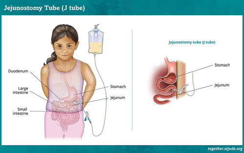
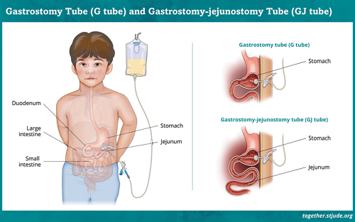
Placement of G Tube, J Tube, and GJ Tube
A gastrostomy is a type of surgery to make a small opening through the skin into the stomach for a feeding tube. The opening is called a stoma. The procedure to create the opening is called an ostomy. Other types of ostomy procedures for feeding tubes include gastrojejunostomy and jejunostomy.
The doctor puts the feeding tube into the stoma. It is called a G-tube (gastrostomy), GJ-tube (gastrojejunostomy), or J-tube (jejunostomy) based on where it is placed. Find information on types of feeding tubes.
Some children have a small plastic device called a button instead of a tube. Your child gets food and medicines through the tube or button.
After the doctor puts the tube in place, a small balloon on the end is filled with water. This keeps the tube from coming out. An external bumper or disc helps hold it in place on the outside of the skin.
A feeding tube ostomy procedure is done 3 main ways:
- Imagery guided
- Surgical (open or laparoscopic)
- Endoscopic (PEG)
The type of procedure your child has can depend on several factors. These may include your child’s health, other planned surgery or procedures, and hospital resources.
Depending on the procedure, you may be told that your child should not eat or drink anything by mouth for a specified amount of time before the procedure. It is very important to follow these instructions, called NPO instructions.
After the procedure, your child will likely stay in the hospital overnight for observation and pain control. Follow your care team’s instructions for tube feeding and giving medicines through a feeding tube. Fluids and tube feedings will be started slowly to make sure that the feeding tube works properly and to help the digestive system adjust.
Risks of feeding tube placement
Placing a feeding tube is a common procedure for children with cancer and other serious illnesses. But there are always risks with anesthesia and surgical procedures. Your doctor will explain the procedure and discuss the risks and benefits.
During the procedure, the main risks are problems with anesthesia or injury to nearby organs. After the feeding tube is placed, the most common problems are:
Serious complications are rare, but they do occur. Follow all care instructions to reduce the risk of infection and keep the feeding tube working properly. Check with your doctor about any changes to medicines after feeding tube placement.
Imagery-guided feeding tube placement
Imagery-guided gastrostomy uses fluoroscopy to guide the placement of the feeding tube through the abdomen and into the stomach. This method is also known as percutaneous radiologic gastrostomy (PRG). In this procedure, a live X-ray of the stomach and abdomen is shown on a video monitor so the radiology team can view the procedure as it is done. The first tube is usually a long tube, but it may be changed for a low-profile device after healing. Sometimes, a low-profile may be placed initially.
Percutaneous radiologic gastrostomy is usually done under general anesthesia. The total time for the procedure is usually about 1-2 hours with anesthesia and recovery.
Surgical feeding tube placement
If your child has surgery to place a feeding tube (G-tube, GJ-tube, or J-tube), it will be done in the operating room under general anesthesia. The feeding tube is inserted directly through the stomach wall into the stomach (G-tube) or small intestine (GJ-tube).
A jejunostomy (J) tube is inserted directly through the wall of the intestine. These tubes are usually low-profile or button devices. If a long tube is placed first, it may be replaced with a low-profile device after the tract heals in about 6 weeks.
The surgery may be minimally invasive (laparoscopic) using several small cuts or incisions. Or it may be an open surgery that uses a larger incision. In laparoscopic surgery, a tiny camera is inserted to guide the procedure. In an open procedure, a larger incision is made through the abdominal wall to reach the stomach or intestine.
When possible, laparoscopic surgery is preferred over open surgery. However, open surgery may be needed if there are medical needs such as scar tissue or disease-related factors. The total time for the procedure is usually about 1-2 hours with anesthesia and recovery.
Percutaneous endoscopic gastrostomy (PEG)
Percutaneous endoscopic gastrostomy (PEG) is the placement of a feeding tube using an endoscope. An endoscope is a thin, long, flexible instrument with a camera and light attached to the end. The endoscope is inserted through the mouth, down the esophagus, and into the stomach.
A video monitor shows a picture of the inside of the stomach. This allows the doctor to place the feeding tube in the correct position. The procedure is usually done under general anesthesia and takes about 1-2 hours with anesthesia and recovery.
The PEG tube is usually a long tube, but it may be changed for a low-profile device after the tract heals.
What to expect after surgery
After the tube is placed, your child will go to the recovery room to wake up from anesthesia. Patients usually stay in the hospital for 1-2 days. While in the hospital, your child will start getting liquids through the feeding tube. Before the formula is started, your child will probably get clear liquids through the tube first.
Your child’s feeding tube will be secured with tape or another device that holds it in place. There might also be gauze around the tube. Nurses will change the gauze and tape in a few days when they clean the area. They might put on fresh gauze and tape as needed. If your child has a device holding the tube in place, it will be changed if it gets dirty or starts to come off.
You might notice a little fluid draining around the tube for 1-2 days after it is put in. After your child has a tube for 6-8 weeks, doctors might change it to a button or smaller tube.
Your child might have some pain after the gastrostomy procedure. If your child has pain, tell your care team. They can prescribe medicines to help. Your child might also get antibiotics to prevent infection.
Your care team will show you how to care for the tube and the skin around it. It is important to keep the tube and your child’s skin clean and free from infection.
You will learn what to do if the tube accidentally falls out or gets plugged. If the tube comes out, it is very important to put it back in as soon as possible. This is because the stoma can close quickly.
A clinical dietitian will be part of your child’s medical team. The dietitian will create a feeding schedule to make sure your child gets enough nutrition. The team will also keep track of your child’s weight.
Your child should be able to return to normal activities after they feel well enough.
Key points about feeding tube placement
- The procedure to create an opening for a feeding tube is called an ostomy. Ostomy procedures for feeding tubes include gastrostomy (G-tube), gastrojejunostomy (GJ-tube), and jejunostomy (J-tube).
- The opening for the feeding tube is called a stoma.
- A feeding tube ostomy procedure is done in 3 main ways: imagery guided (PRG), surgical (open or laparoscopic), and endoscopic (PEG).
- Your care team will show you how to give feedings, how to care for the feeding tube and the skin around it, and what to do if the feeding tube comes out.
- Your child should be able to return to most normal activities after they feel well enough.
-
Skin Care for Feeding Tube Sites
If your child has a feeding tube, such as an NG or G tube, it is important to take care of the skin around the tube. Learn skin care tips for feeding tube sites.
-
Feeding Tube Placement
Placement of a feeding tube into the stomach or intestine is a common procedure in children with cancer or other illnesses. Learn about feeding tube placement.
-
Placement of NG Tube and NJ Tube
Placement of a feeding tube through the nose is a common procedure in children with cancer. A feeding tube may be placed into the stomach (nasogastric or NG tube) or into the intestine (nasojejunal or NJ tube).


