Ultrasound imaging is also called sonography. It uses high-frequency sound waves to create images that show the inside of the body. These sound waves cannot be heard by the human ear.
Ultrasound images show what is happening in the body at that moment. They can show the structure and movement of the body’s internal organs. They can also show how blood flows through the blood vessels.
Ultrasound imaging is a safe way to clearly see tissues and organs inside the body.
How does ultrasound work?
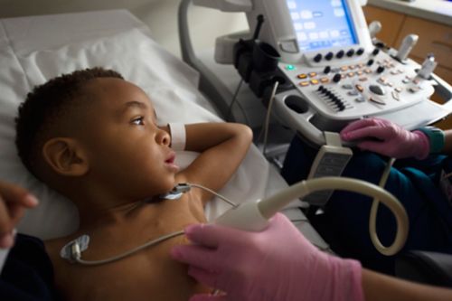
An ultrasound technologist uses a probe called a transducer to see the inside of the body. A transducer sends sound waves into the body. The waves are reflected by organs and tissues. This produces an image of the internal organs.
- For the ultrasound exam, your child lies down on an exam table. The area that will be scanned is uncovered.
- A technologist puts clear, water-based gel on the area of the body that will be scanned. This gel helps the transducer make better contact with the skin. The gel might feel cold at first.
- Next, the technologist presses the transducer against the skin on the area to be scanned. To get the best image, the technologist moves the transducer across that area.
- When the ultrasound is over, the technologist cleans the gel off your child’s body.
Some ultrasound exams use a contrast agent. The contrast helps your provider see the blood flow in the organs in the body.
Contrast is injected into the blood. If your child does not already have an IV or port, then the nurse will need to start an IV. If an IV is needed, the staff will talk to you and your child before starting the IV.
The contrast agent that is used is a type of microbubbles. These are small particles about the same size as a red blood cell. The microbubbles can be seen very well on ultrasound imaging. This is a safe, effective way to enhance an ultrasound image.
The contrast agent looks milky. It comes in a small vial. Side effects are rare and do not last long. If you have questions about the use of microbubbles, ask a member of your care team.
A Doppler ultrasound study may be a part of an ultrasound exam. Doppler ultrasound can show blood flowing through blood vessels, including the body’s major arteries and veins. A Doppler ultrasound can help show these kinds of features:
- Objects blocking blood flow, such as clots.
- Blood vessels becoming narrow.
- Tumors and issues in the blood vessels and vascular system.
Ultrasound images are seen as they are happening.
Ultrasound uses
Diagnostic ultrasound
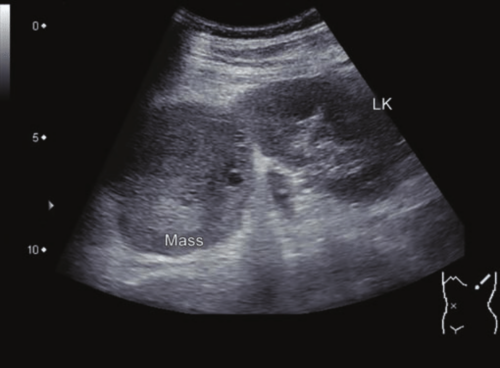
Ultrasound can aid in the diagnosis of pediatric cancers, including:
Ultrasound can also be used to guide needle biopsies . These biopsies help diagnose tumors. Ultrasound can help doctors see how the tumor is involvement with nearby blood vessels.
Ultrasound imaging is used to look at the:
- Appendix
- Stomach/pylorus
- Liver
- Gallbladder
- Spleen
- Pancreas
- Intestines/colon
- Kidneys
- Bladder
- Testicles
- Ovaries
- Uterus
- Thyroid
- Arteries and veins
Ultrasound screening
Some pediatric facilities use ultrasound to screen a patient with a genetic condition. The genetic condition can make them more likely to develop tumors. For example, patients with Beckwith-Wiedemann, Denys-Drash, Frasier, and WAGR syndromes have an increased chance of developing Wilms tumor.
A transducer sends sound waves into the body. Organs and tissues reflect those waves. This reflection allows a picture to be made of organs inside the body.
How to prepare for your child's ultrasound
- Make sure your child wears comfortable, loose-fitting clothing. They may need to remove certain clothing or jewelry in the area of the body that will be examined. Or they may need to wear a hospital gown during the exam.
- For an ultrasound of the liver, gallbladder, spleen, and pancreas, your child should not eat or drink for 6 to 8 hours before the test.
- For ultrasound of the kidneys, bladder and pelvis, your child may be asked to drink up to 64 ounces of liquid 2 to 3 hours (without urinating) before the test to fill the bladder. The best liquid to drink is water. It might be hard to get a small child to drink 64 ounces, but the goal is to drink as much as possible without emptying the bladder.
Follow the instructions of your care team to make sure your child is ready for the ultrasound.
A radiologist, a doctor trained to read radiology exams, looks carefully at the images. Then that doctor sends a report to the care team member who asked for the exam. The primary doctor will share the results with you and your child. In rare cases, the radiologist may talk about the results at the end of your child’s exam.
A follow-up exam may be needed so that doctors can watch issues or changes over time. Follow-up exams are sometimes the best way to see if treatment works or if an issue is stable over time.
—
Reviewed: September 2022


