Skip to Main Content

Welcome to

Together is a new resource for anyone affected by pediatric cancer - patients and their parents, family members, and friends.

Learn More
Blog

Laboratory Tests

A patient’s medical team uses the specific, detailed information from laboratory testing to create a treatment plan for that individual case.

Blood cancers such as leukemia are diagnosed by examining blood and bone marrow. Other types of cancer are typically diagnosed by examining tissue removed from a suspected tumor during a biopsy. The sample of tissue removed for testing is called a specimen.

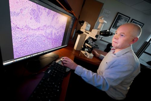
A pathologist reviews an enlarged view of tissue on a large format monitor.

Pathologists specialize in different areas. It is important that specimens are examined by pathologists who have specialized training for the specific cancer type they are evaluating.

These tests take place in a pathology lab, led by a pathologist, a medical doctor who specializes in diagnosing disease by studying cells, tissues, and body fluids. Like other medical doctors, pathologists specialize in different areas.

  • Hematopathologists focus on diagnosing cancers of the blood, such as leukemias and lymphomas.
  • Anatomic pathologists study body organs and tissues. In cancer diagnosis, anatomic pathologists focus on types of tumors such as sarcomas and carcinomas.
  • Neuropathologists are a special type of anatomic pathologist that focus on diagnosing cancers of the brain and spinal cord.

It is important that specimens are examined by pathologists who have specialized training for the specific cancer type they are evaluating.

How specimens are collected

Cancers of the blood

In cases of suspected cancer of the blood, a sample of blood or bone marrow (through a bone marrow aspiration and/or biopsy) will be collected from the patient. The staff labels the container with the patient’s information, and the specimen is sent to a pathology laboratory.

Tumors of solid tissue

When a patient has a suspected tumor in the brain or other parts of the body, all or part of it may be removed during surgery. When only a portion of the tumor is removed and evaluated, this is called a surgical biopsy. In other surgical procedures, surgeons remove as much of the tumor as possible. This is referred to as a resection.

The removed tissue must be cut into thin sections, placed on slides, and stained with dyes before it can be examined under a microscope. Two methods are used to make the tissue firm enough to cut into thin sections:

  • Frozen section - Tissue is rapidly frozen.
  • Permanent section - Tissue is embedded in a special wax called paraffin.

All tissue samples are prepared as permanent sections, but sometimes frozen sections are also prepared.

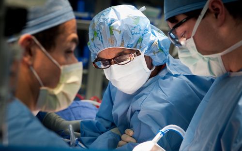
In surgical biopsies, the staff may prepare a frozen section so a pathologist can analyze the biopsy specimen during the surgery. The specimen is frozen quickly and prepared so a pathologist can examine it in a laboratory close to the operating suite. The quality is not as high as with a permanent section, but the pathologist can usually quickly determine if the tissue is cancerous. This knowledge helps surgeons make immediate decisions about surgery.

In some cases of suspected tumors of solid tissue, another type of biopsy – a needle biopsy – is performed. In a needle biopsy, a sample of tissue is removed with a needle. A member of the team who performed the biopsy will place the specimen in a sterile container and a special liquid will be added to preserve it. It will be labeled with the patient’s information and sent to the pathology laboratory for examination and testing.

What happens in the laboratory

The pathology lab staff prepares the sample and performs different types of tests to gather information about the tissue. The pathologist includes this information in a report for the oncologist. The pathologist and oncologist work together to make a diagnosis.

Gross examination

First, a pathologist will examine the specimen and describe its appearance to the naked eye. This is called a gross examination. This description will include the specimen’s color, size, and other features.

Preparation of specimen

The laboratory staff then prepares the specimen for the pathologist to look at under a microscope. The specimen is processed in a machine that places the tissue in a paraffin wax block. The staff cuts the block of tissue into thin slices to mount on slides (tissue sections). Slides are small, flat pieces of glass used to view objects under a microscope.

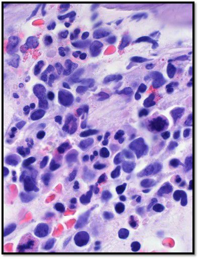
After this process, a technician will stain the tissue section with dyes (typically with hematoxylin and eosin) which help highlight different features of the tissue under the microscope. The nucleus of each cell will appear blue. The rest of the cell (cytoplasm) will appear pink.

Sample histology slide shows tissues stained so that the nucleus of the cells appear blue and the rest of the cell appears pink.

Sample histology slide shows tissue stained with hematoxylin and eosin to highlight different features of the tissue under the microscope.

Microscopic examination

The pathologist looks at the tissue sections under a microscope to see how they compare with normal cells. Viewing the specimen in this manner is called histology. Histology is the study of structures of cells and tissue.

The pathologist creates the report based on what he or she sees under the microscope and the results of other tests performed on the tissue. It is written in technical, medical language that may be hard to understand. Ask your care team if you have any questions.

In general, the pathologist describes the specimen’s features:

  • The types of cells
  • How the cells are arranged
  • Whether the cells are cancerous
  • Other features helpful in diagnosis and treatment such as whether the tumor cells demonstrate aggressive features (often referred to as tumor grade) or if the tumor cells have spread into other normal tissues (often referred to as the tumor stage). In some circumstances, pathologists also determine if the tumor has been completely removed from the patient or if the edge of the specimen (margins) are positive for cancer.

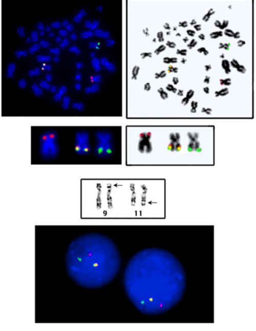
In addition to evaluation under the microscope, the specimen may undergo further tests and analysis. These results will also be included in the pathology report.

Finding out the results

Sometimes all testing and analysis happen at the medical center where the biopsy was performed. Other times, the specimen must be sent to laboratories located somewhere else. Some tests require a series of steps and procedures and may take a few weeks for results.

The pathologist will include information from these tests into the pathology report. It is shared with the oncologist. The oncologist will share results of the pathology report during a clinic visit.

The more information doctors have about the cancer the better they can develop the most effective therapies for that individual case of cancer.


Reviewed: July 2018