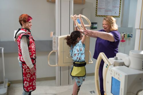
An x-ray is a type of imaging test that creates black-and-white pictures of the inside the body. Health care providers use x-rays to help diagnose and monitor injuries or illnesses.
An x-ray machine sends a small amount of radiation to different parts of the body.
The shades of black and white in an x-ray image depend on how much radiation passes through the body:
- Dense structures such as bones and metal look white because they absorb the most radiation.
- Fat and other soft tissues look gray because they absorb less radiation.
- Air absorbs the least radiation. Lungs and other structures that contain air look black.
X-rays are quick and painless. They can provide important details about your child’s health. An x-ray can help your care team see what is going on inside the body. X-rays can show:
- Evidence of cancer or other health problem
- Infections
- Bone fractures or other problems in bones or joints
- Tooth cavities
- Foreign objects in the body
An x-ray is not painful. But your child needs to stay still so that the image is clear. Talk with your child about the importance of not moving during the test.
A health care professional called a technologist will take your child’s x-rays. Be sure to let the technologist know if your child is nervous or uncomfortable.
You may be allowed to be in the room while x-rays are taken. Let staff know if anyone who will be in the x-ray room (patient or caregiver) might be pregnant.
Your child may be asked to remove jewelry, watches, or glasses. They may need to wear a hospital gown.
For some x-rays, a contrast medium—such as iodine or barium—is used to give better details on the images. If your child has ever had a reaction to an x-ray contrast, tell the technologist or a member of your care team.
What to expect during an x-ray
The x-ray technologist will help your child get position. Your child may stand, sit, or lie down. The position will depend on the machine and the area of the body being x-rayed.
It is important to get the clearest view possible. Sometimes, special padding or straps are used to keep the child still. Your child may need to hold their breath because moving will blur the x-ray.
The technologist will step behind a protective barrier and ask your child to stay very still for a few seconds while the x-ray is taken.
The technologist may help your child change position if more x-rays are needed.
If your child wore a gown during the x-ray, they can change back into their clothes when the x-ray is done.
After the x-ray the images will be sent to a radiologist. This doctor specializes in interpreting imaging tests. The radiologist will look at the x-ray and send a report to your health care provider.
Possible risks of an x-ray
X-rays use a very small amount of radiation to take images. This is safe for most people. But it is important not to have too many x-rays unless your health care provider says they are needed. The doctor will only order x-rays when necessary. People are exposed to radiation every day in the environment. The medical benefits of x-rays outweigh the small amount of radiation exposure.
Questions to ask your care team


